Digestive and Excretory Systems for the DAT
/Learn key DAT concepts related to GI anatomy, digestion, absorption, nephrons, and endocrine control, plus practice questions and answers
Table of Contents
Part 1: Introduction to the digestive and excretory systems
Part 2: Digestive system
a) Gastrointestinal anatomy
b) Accessory organs
c) Enzymatic digestion and absorption
Part 3: Excretory system
a) Urine production
b) The nephron
c) Endocrine control
Part 4: High-yield terms
Part 5: Questions and answers
----
Part 1: Introduction to the Immune System
Energy plays a large role in your body’s ability to maintain homeostasis and function. Both the digestive and excretory systems collaborate to process ingested food into energy and expel waste from the body. The DAT often assesses concepts related to these systems, including aspects of kidney function and structure. A solid understanding of these mechanisms is invaluable, particularly for success on the DAT.
Key terms critical for comprehension are bolded throughout this guide. Additionally, a set of questions styled after the DAT offer you an opportunity to assess your knowledge.
----
Part 2: Digestive system
a) Gastrointestinal anatomy
The human body houses a lengthy, tube-like structure known as the digestive tract, spanning the torso and abdomen. As food enters this tract through the mouth, it undergoes a sequence of digestive steps to extract essential nutrients, while unabsorbed nutrients are expelled as waste.
The process of digestion initiates in the mouth, where both mechanical and chemical breakdown occurs. Chewing, termed mastication, is the primary mechanical digestion method in the mouth. Mastication breaks down food into smaller particles, thereby increasing the surface area. Salivary enzymes, including salivary amylase and salivary lipase, commence the chemical digestion of sugars and lipids within the food. Food only spends a brief amount of time in the mouth, so digestion here is limited and the process continues further along the digestive tract.
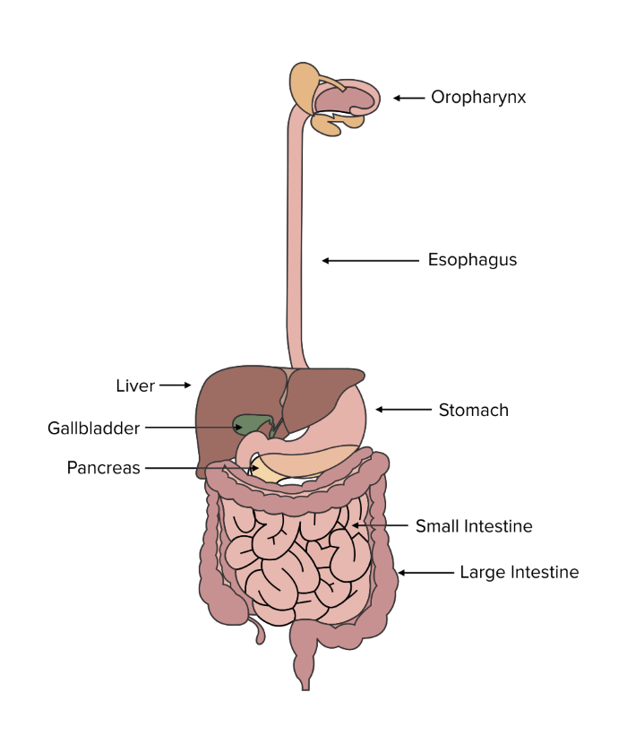
FIGURE 1: ANATOMY OF THE DIGESTIVE TRACT
Before swallowing, the tongue molds food into a ball-shaped mass termed a bolus. Muscles facilitate the movement of the bolus from the mouth to the oropharynx.
Swallowing guides food through the pharynx, a tube shared by the respiratory and digestive systems. To prevent food from entering the lungs, the epiglottis—a flap-like structure—instinctively seals the opening of the larynx. This allows food to safely travel through the pharynx and enter the esophagus.
Linking the mouth to the stomach, the esophagus features upper and lower esophageal sphincters. These sphincters are ring-shaped muscles that regulate food entry into the esophagus and stomach.
The upper third of the esophagus is controlled by skeletal muscles that allow you to swallow. Smooth muscles control the rest of the esophagus and propel food further down the digestive tract through wave-like, involuntary contractions. This process of movement is called peristalsis and is made easier by saliva that has mixed with the bolus.
Continuing its journey, food reaches the stomach, a pivotal site for chemical digestion. Glands within the stomach lining secrete acidic chemicals and enzymes essential for digestion. These are the digestive cells and enzymes that you need to know:
- Parietal cells release hydrogen ions.
- Chief cells secrete pepsinogen, a precursor to the enzyme pepsin that breaks down proteins.
- G-cells secrete gastrin, which stimulates hydrochloric acid secretion, establishing an acidic environment.
- Goblet cells secrete mucus, protecting the stomach lining from acidic damage.
With the aid of muscular contractions, enzymes, and acid within the stomach, food transforms into chyme. Chyme is a thick mixture that progresses to the small intestine for further digestion and absorption of nutrients.
The small intestine, comprising the duodenum, jejunum, and ileum, facilitates nutrient digestion and absorption. Alkaline juices from the pancreas neutralize acidic stomach contents, initiating enzyme release. Microvilli in the small intestine enhance surface area and aid in nutrient absorption, which is primarily facilitated by diffusion and active transport into capillaries.
For hydrophobic molecules, such as fat-soluble vitamins K, A, D, and E, lacteals facilitate entry into the lymphatic system, aiding absorption.
The large intestine houses beneficial gut flora that assist in nutrient production and absorption, particularly vitamin K. Water and salts are also absorbed within the large intestine. Eventually, waste material accumulates as fecal matter and is stored before being expelled through the rectum and anal sphincters.
The enteric nervous system, part of the autonomic nervous system, influences digestion by governing movement and peristalsis within the digestive tract.
b) Accessory organs
Although not physically part of the tubular digestive tract, the liver, gallbladder, and pancreas significantly contribute to chemical digestion.
Positioned in the upper-right abdomen, the liver serves multifaceted roles in digestion, notably producing bile. This bile, comprising salts, cholesterol, and bilirubin pigment, aids fat digestion. Additionally, the liver produces albumin, facilitating hormone transport in the bloodstream. Moreover, it detoxifies the blood, stores or converts excess nutrients like glycogen and triacylglycerols, and provides energy through processes such as gluconeogenesis and glycogenolysis.
Underneath the liver, the gallbladder acts as a bile reservoir, releasing bile into the small intestine upon stimulation by the hormone cholecystokinin. Bile doesn't chemically digest fats but helps emulsify them, enabling better mixing with digestive juices.
The pancreas secretes enzymes—pancreatic amylase, peptidases, and lipases—essential for digesting carbohydrates, proteins, and lipids. These enzymes reach the duodenum via the pancreatic duct system, produced and released by acinar cells.
These organs are exocrine, meaning they release substances externally rather than internally like endocrine glands. They secrete substances into the digestive tract, analogous to sweat or sebaceous glands that secrete substances onto the skin.
Remarkably, the pancreas is both exocrine and endocrine. Beyond digestive juices, it produces insulin to regulate blood sugar levels. While its role in insulin production is pivotal, the pancreas has broader functions in the endocrine system, detailed in our endocrine system guide.
c) Enzymatic digestion
Enzymes are pivotal in the digestive process, especially in the stomach where pepsin aids in breaking amino acid bonds and enhancing food surface area. Further enzymatic digestion occurs in the duodenum, where accessory organs release additional enzymes.
Within the duodenum, enzymes play a crucial role in chyme digestion. Brush-border enzymes break down carbohydrate chains into absorbable monomers. Notably, brush-border disaccharidases like maltase, lactase, and sucrase split maltose, lactose, and sucrose into monomers. Peptidases, including dipeptidases and aminopeptidases, work on proteins by breaking down dipeptides and removing N-terminal peptide chains.
Among the duodenum enzymes, enteropeptidase stands out. It activates trypsinogen into trypsin, a vital step in triggering pancreatic enzyme activation within the duodenum.
The duodenum also relies on two key hormones—secretin and cholecystokinin—that are triggered by chyme presence. These hormones facilitate the release of bile and enzymes from the gallbladder and pancreas. Without their action, critical enzymes like pancreatic amylase and peptidases wouldn’t be secreted. Pancreatic amylase is responsible for polysaccharide digestion into disaccharides, and pancreatic peptidases (trypsin, chymotrypsin, and carboxypeptidases A and B) are responsible for protein breakdown. Additionally, pancreatic lipase, vital for fat digestion, relies on these hormones for secretion.
| |
|
|
Carboxypeptidases A and B |
||
Pancreatic amylase |
||

Gain instant access to the most digestible and comprehensive DAT content resources available. Subscribe today to lock in the current investments, which will be increasing in the future for new subscribers.